
Morphology & Imaging Core
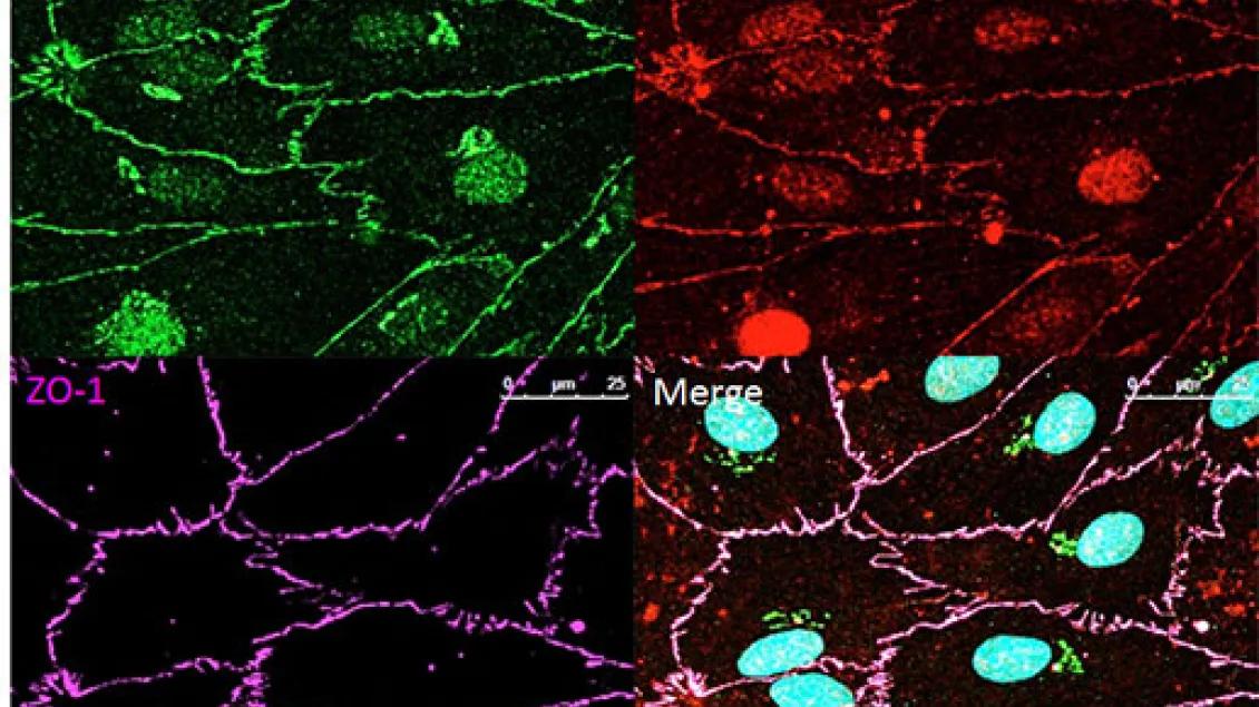
The Morphology & Imaging Core in the U-M Medical School Department of Ophthalmology & Visual Sciences provides specialized histology services for vision researchers, focusing on the processing of ocular and brain tissues. We support investigators through expert sample preparation, publication-quality imaging assistance and technical training.
For More Information
Project Priorities
Comprehensive Tissue Processing
Imaging Analysis & Publication Support
Technical Training & Consultation
Our Services
- Process ocular and brain tissues for light microscopy, immunocytochemistry and in situ hybridization.
- Prepare and fix specimens for electron microscopy to be analyzed at other facilities.
- Provide training in tissue processing, microtomy, staining, and immunohistological techniques.
- Assist with image analysis and the production of publication-quality figures.
Contact
Brad Nelson, Manager/Technician
Email: [email protected]
Phone: 734-615-7991
Fax: 734-936-3815
Instrumentation
Note: Some pieces of equipment must be reserved for use. Reserve equipment
- Cryostar NX50 Cryostat
- Leica CM3050 Cryostat
- Lecia CM3050S disposable blade cryostat
- Reichert steel knife sharpener
- Leitz rotary microtome for plastic sectioning
- Tissue-Tek Paraffin processing and embedding station
- Access to Shandon AS 325 paraffin microtome
- Shandon Tissue Processor Embedding Station
- Standard light level biological stains and staining setup available
- Two research-quality Leica DM 6000 microscopes with 2X-100X objectives, differential interference contrast optics (DIC), and transmitted and epi-fluorescence light sources. The microscope is fitted with duel digital cameras (greyscale for fluorescence, color for high resolution color stain) and interfaced with an HP computer with Leica’s LAS-X for image capture and analysis.
- Epson Expression 1680 Flatbed scanner with dedicated G5 iMac running Adobe Creative suite (Adobe Photoshop, Illustrator, In-Design with PageMaker plug-in, Acrobat and Go-Live). Also, Microsoft Office X, and OCR software
- Nikon Super Coolscan 5000ED slide scanner, also associated with a G5 iMac
- Access to the UM Microscopy and Image Analysis Laboratory (MIAL) in the Department of Cell and Developmental Biology for use of a BioRad or Zeiss laser scanning confocal microscope and a Philips transmission electron microscope
- Olympus EX51 Microscope
- Leica 205FA Fluorecence Stereo Microscope
- Leica MetaMorph & LAS-AF Offline Imaging Workstation
- Leica SP5 Confocal Microscope
- Leica SPX5 2-Photon Laser-Scanning Confocal Microscope
- LMD7000 Laser Micro dissection Microscope
- Ophthalmology Antibody Inventory (access limited to UM Vision Research Community)
Equipment Reservation Calendars
Note: Log into the MiCores reservation calendar site to view these calendars.
Core Director
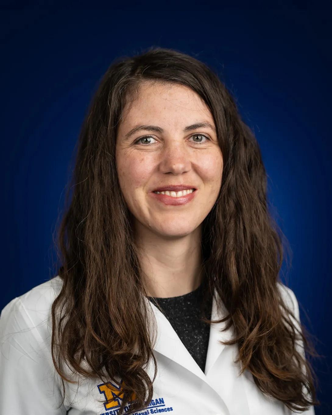
Jillian N Pearring, PhD
Skillman Early Career Professor of Pediatric Ophthalmology
Associate Professor of Ophthalmology and Visual Sciences
Associate Professor of Cell and Developmental Biology
Medical School
Associate Professor of Ophthalmology and Visual Sciences
Associate Professor of Cell and Developmental Biology
Medical School
