Neurosurgery Resident Research
Uniquely, residents in the U-M Neurosurgery Residency Program dedicate PGY-4 and PGY-5 years to research and innovation. Residents plan for and design their research projects in advance. While research is typically performed on campus, a number of residents have pursued funded projects at other institutions.
For More Information
Resident Research Profiles
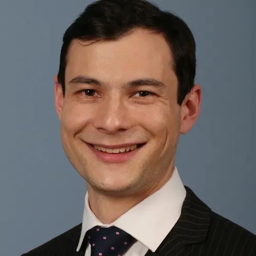
Dr. Jordan Lam’s research with Dr. Cynthia Chestek and Dr. Matthew Willsey focused on developing next-generation brain-computer interfaces to restore function for patients with neurological disorders.
Dr. Lam’s work bridged the translational spectrum and scientific disciplines, and integrated mechanical testing, immunohistochemistry, and electrophysiological recording in various animal models. Notable projects included the development and manufacture of subcellular carbon fiber electrodes and the first-in-human implantation of the novel Paradromics Connexus electrode array. Dr. Lam’s research was supported by the NIH Ruth L. Kirschstein National Research Service Award (T32), the Coulter Translational Research Program, and the Marcus Foundation.
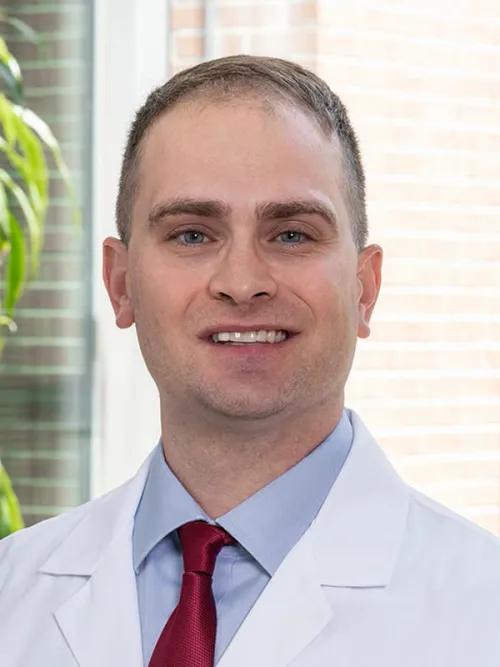
Dr. Ward’s clinical focus is on complex spine, peripheral nerve, and brachial plexus surgery. His research priorities included brain computer interfaces, neuroprosthetics, and molecular basis of nerve regeneration.
During his dedicated research time during R4 and R5, he focused on nerve regeneration and brain-machine interfaces to restore limb function. In the lab of Dr. Cynthia Chestek, he led a large animal project that developed and optimized a novel peripheral nerve interface system to improve dexterity and fatigue in the upper extremity after spinal cord injury. In the lab of Dr. Roman Giger, he co-led several projects to explore immune-based regeneration in the peripheral nervous system, utilizing single-cell RNA sequencing to investigate molecular differences between regenerating infant and adult nerves. For this work he was awarded a $150,000 grant from the Merkin Peripheral Nerve/Regeneration Center at Johns Hopkins University, with a goal to find new molecular targets to jump start PNS and CNS nerve regeneration in patients.
The final phase of Dr. Ward’s research program is to integrate these technologies into a multifaceted approach to nervous system injury. In the context of CNS/PNS trauma, he works to integrate these domains into restorative therapies and solutions for patients with devastating neurological injury and disease.
Matthew Willsey, MD, PhD completed a PhD in Biomedical Engineering during his 2-year protected research time with an additional year-long leave-of-absence. His dissertation research with Drs. Parag Patil and Cynthia Chestek focused on Restorative Neuroengineering, including intraoperative modulation of sensorimotor pathways, the effects of anesthetics on cortical signal flow, and brain-machine interface neuroprosthetics.
To support this research, Matt received an NIH T32 training award and funding from a new $2.3M brain machine interface grant from the NSF to the Restorative Neuroengineering Group at the University of Michigan, which is headed by Dr. Parag Patil.
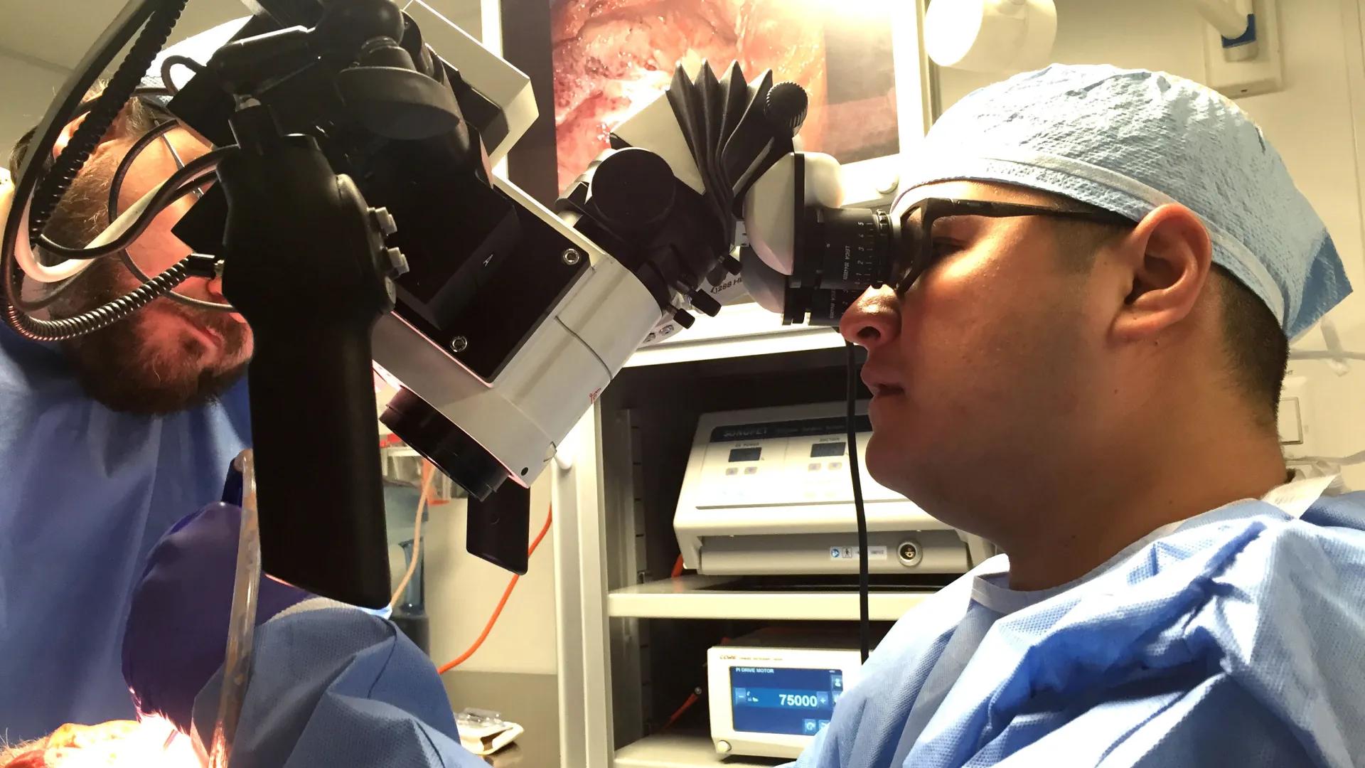
The primary focus of Todd Hollon's research program was to combine stimulated Raman scattering (SRS) microscopy, a label-free, live-cell, optical imaging modality, with modern deep learning and computer vision techniques to discover the molecular, cellular, and microanatomic features of skull base and malignant brain tumors.
This work aimed to:
- Improve the use of histologic data in neurosurgical oncology
- Develop novel deep learning algorithms to improve computer vision systems in microscopy
- Explore basic science applications towards characterizing diseased states and monitoring treatment responses. Current intraoperative pathology workflow is cumbersome, time-intensive, depletes scant specimens, and prone to artifact.
These limitations severely limit the neurosurgeon’s ability to evaluate tissue in the operating room. Due to rapid digital image capture and rich histochemical information in SRS images, they have used neural network-based computer vision systems to automate the interpretation of fresh pediatric and adult brain tumor specimens in an order of magnitude less time (~2 mins). Additionally, they have collaborated with other neuropathology investigators to use SRH to monitor disease burden and treatment response in lysosomal storage diseases both in vitro and in mouse models. The final phase of Dr. Hollon's research program focused on using SRS microscopy to intraoperatively identifying molecular driver mutations and metabolic aberrations in brain tumors, especially gliomas (e.g. IDH-1/2, 1p19q, MGMT methylation) and meningiomas (e.g. NF-2, SMO, AKT).
Luis E. Savastano's research was focused in the development of new technologies to better understand, diagnose and treat cerebrovascular diseases. He worked with Tom Wang and Eric Seibel at the University of Michigan to develop a new intravascular laser-based angioscope for structural and biological images of atherosclerosis.
This work received the Cerebrovascular Research Award of the American Association of Neurological Surgeons, the Galbraith Award of the Congress of Neurological Surgeons and was presented twice at the Academy. In addition, he worked with Albert Shih at the University of Michigan in a NIH-funded project to develop a new platform for mechanical thrombectomy in stroke.
Shawn Hervey-Jumper, MD examined the role of microRNAs in brain tumors. He worked with Dr. Xing Fan in the Crosby Neurosurgical Laboratories at the University of Michigan. His work was supported by a T32 training grant from the NIH (Cancer Biology Training Program).
Arnold B. Etame, MD was a Neuro-Oncology Research Fellow at the Labatt Brain Tumor Research Center at the Sick Children’s Hospital in Toronto and is a Ph.D. candidate in the Department of Laboratory Medicine and Pathobiology at the University of Toronto.
He worked with Dr. James Rutka. His research was focused on using nanoparticles to target brain tumor infiltration. He received the Congress of Neurological Surgeon Wilder Penfield Fellowship as well as a Fellowship from the University of Toronto.
Daniel Orringer's research focused on nanodevices designed to visibly delineate tumor margins in the operating room to enhance the precision of the brain tumor surgery.
The research was performed in collaboration with Dr. Kopelman in the Department of Chemistry and Dr. Oren Sagher at the University of Michigan. The work was supported by a Ruth L. Kirschstein National Research Service Award from the National Cancer Institute and the Congress of Neurological Surgeons Basic/Translational Research Fellowship.
Dr. Arushi Tripathy's clinical interests include complex brain tumor surgery and the use of intraoperative mapping. Her research focuses on treatment resistance in primary brain tumors, central nervous system liquid biopsy, and anti-thrombotic therapy management for patients with hemorrhagic brain tumors. During her dedicated research time working with Drs. Wajd Al-Holou, Daniel Wahl, and Carl Koschmann, she developed an in vitro model of treatment resistance in glioblastoma and performed comparative sequencing and mechanistic studies to elucidate pharmacologically targetable pathways. Dr. Tripathy has also studied longitudinal liquid biopsy biomarkers in primary and metastatic tumors, coordinated a clinical trial for surgical site reservoir placement at the time of glioma tumor resection for longitudinal peri-tumoral cerebrospinal fluid sampling, and examined national practice patterns and outcomes in hemorrhagic brain tumors to propose guidelines for anti-thrombotic therapy management and inform future areas of investigation.
Dr. Tripathy's work has been supported by the Neurosurgery Research Education Foundation Andrew T. Parsa Research Fellowship, an NIH T32 Award for Training in Clinical and Basic Neuroscience, and by a University of Michigan Rogel Cancer Center Discovery Award.
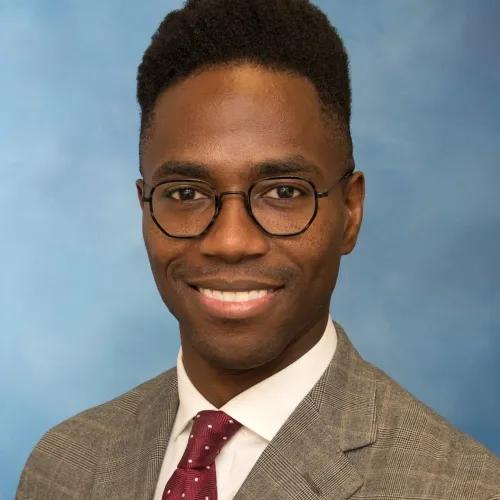
Dr. Edwin Nieblas-Bedolla’s clinical interests within neurological surgery encompass the treatment of skull base and cerebrovascular disease. His research interests are broad, with an emphasis on understanding how cancer interacts with the nervous system, and leveraging these insights to develop novel therapeutic strategies.
During his training, Dr. Nieblas-Bedolla has contributed to our knowledge of molecular and cellular mechanisms underlying primary and metastatic brain tumor behavior, performed translational studies evaluating potential anti-cancer drugs, and involved in the management of a clinical trial assessing an iron-chelating drug in aneurysmal subarachnoid hemorrhage. He has also studied healthcare disparities, and continues to be involved in research surrounding equity in medicine. He is pursuing a PhD in Neuroscience at the University of Michigan during his neurosurgery training. Dr. Nieblas-Bedolla’s work has been supported by an NIH T32 Early Stage Training in the Neurosciences Training Grant.
Recent Resident Publications
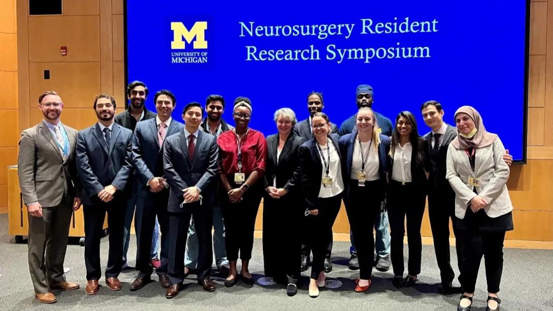
Neurosurgery Resident Research Symposium
Since 2014, the annual U-M Neurosurgery Resident Research Symposium at the U-M Medical School has promoted the academic productivity of our residents and brought together research and clinical colleagues in neurosurgery.
This daylong symposium supports our educational mission of training exemplary neurosurgeons who have a strong background in clinical neurosurgery and research. Each spring, residents present their research, and awards are given to the best clinical and basic science presentations. Additionally, a nationally recognized neurosurgeon who is also a Visiting Professor, is invited to deliver the keynote address and help judge presentations.
The Chandler Clinical Research Award is presented for the best clinical research presentation, and the Crosby Basic Science Award is given to the best basic science presentation.
Past Keynote Speakers & Award Winners
- Dr. Frederick Lang (University of Texas MD Anderson Cancer Center)
- Elizabeth Crosby Visiting Professor
- "The perivascular niche and mesenchymal stem cells in gliomas"
- Crosby Basic Science Award: Ayobami Ward
- Chandler Clinical Research Award: Katherine Holste
- Dr. Rajiv Midha (University of Calgary)
- Elizabeth Crosby Visiting Professor
- "Advances in nerve repair: experimental to clinical"
- Crosby Basic Science Award: Ayobami Ward
- Chandler Clinical Research Award: Sravanthi Koduri
- Dr. Jennifer Strahle (Washington University)
- Joan Venes Visiting Professor
- "The CSF-brain axis: insights into pediatric neurosurgical diseases"
- Crosby Basic Science Award: Katherine Holste
- Chandler Clinical Research Award: Sravanthi Koduri
- Amy Heimberger (Northwestern University)
- Elizabeth Crosby Visiting Professor
- "Immune reactivity in the CNS tumor landscape -- implications for immunotherapy"
- Crosby Basic Science Award: Todd Hollon
- Chandler Clinical Research Award: Junior Daou
Resident Research Symposium Highlights
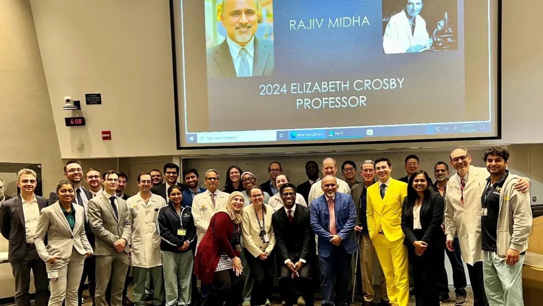
Neurosurgery faculty, residents, and Dr. Midha at the 2024 event.
Residents and Dr. Heimberger (center) at the 2022 event
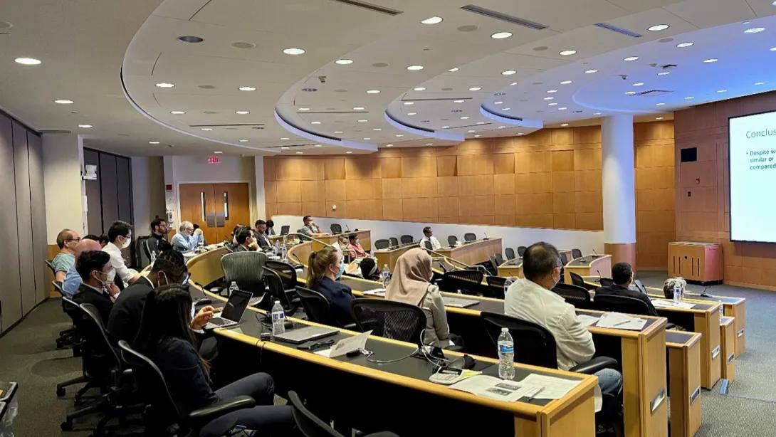
Members of the audience at 2022 event in the Danto Auditorium.