
Nicole Seiberlich, PhD
Manage Your Profile
About
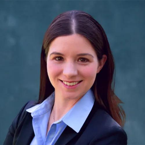
Dr. Nicole Seiberlich is currently the endowed Research Professor of Cardiovascular Imaging and a Full Professor (with tenure) in the Departments of Radiology and Internal Medicine (Cardiology) at the University of Michigan. Prior to this role, she served as the endowed Elmer Lincoln Lindseth Professor of Biomedical Engineering at Case Western Reserve University. She is the initiator and Co-Director of the Michigan Institute of Imaging Technology and Translation (MIITT) in the Department of Radiology at the University of Michigan. The purpose of this group is to bring together experts in clinical radiology and MRI research to develop and deploy novel MRI techniques for clinical applications. She has grown the group from an initial 8 members in 2019 to more than 35 trainees and scientists, and has developed the research infrastructure required for translational research (including bringing in two MRI systems, hiring technologists and clinical coordinators, and developing a sustainable financial plan). Dr. Seiberlich received the University of Michigan “Technology and Innovation Award” in 2023 for her success in initiating and developing MIITT. Additionally, Dr. Seiberlich is deeply involved in the leadership of multiple international societies which dictate the direction and focus of Magnetic Resonance Imaging in clinical practice. She has served on the Board of Trustees for both the International Society for Magnetic Resonance in Medicine (ISMRM) and the Society for Cardiovascular Magnetic Resonance (SCMR). Additionally, she organized the Annual Meeting of the ISMRM as the Program Chair in 2021, and has been elected by the membership to serve as President of the ISMRM from 2027-2028.
Links
MIITT
Qualifications
-
Post-Doctoral FellowUniversity Hospitals Case Medical Center, Radiology, Cleveland, United States
2008 - 2010
Postdoctoral Research
-
PhDUniversity of Würzburg, Würzburg, Germany
2003 - 2008
-
BSYale University, New Haven, Connecticut, United States
1997 - 2001
Center Memberships
-
Center MemberSamuel and Jean Frankel Cardiovascular Center
Research Overview
Cardiovascular Imaging
Improved Diagnosis using Magnetic Resonance Imaging
Low-Field MRI for World-Wide MRI
Real-time Cardiac MRI
Quantitative MRI
Dr. Seiberlich’s research focuses on the development and translation of rapid and quantitative Magnetic Resonance Imaging (MRI), with applications in the heart, liver, prostate, and brain. This research involves designing both specialized MRI acquisitions as well as image reconstruction techniques, and requires a deep understanding of MRI physics, signal processing methods, the needs of imaging physicians and radiologists, and industrial collaboration. MRI is able to non-invasively probe the structure and function of the human body, and is used extensively in hospitals for the diagnosis and follow-up of disease. Because diseased tissues and healthy tissues have different properties, images made which highlight or measure these properties can be used to identify pathology. However, MRI acquisitions are long, in particular if quantitative measurements of tissue properties are required for a diagnosis. The slow data acquisition results in a number of negative consequences: for example, images of organs which experience rapid and irregular motion such as the arrhythmic heart can be corrupted, and limited data with reduced accuracy and precision are collected for tissue property mapping. Dr. Seiberlich has invented a number of technologies to accelerate the collection of MRI images and accurate quantitative maps. These methods have been demonstrated for improved imaging of the heart and liver—both of which experience substantial motion, as well as accurate tissue property mapping in multiple organs throughout the body. These technologies have led to 26 United States Patents (with another two pending), and six foreign patents (in China, South Korea, Japan, France, and Germany), all of which have been non-exclusively licensed by Siemens Healthineers.
The most well-developed technique developed by Dr. Seiberlich is known as Magnetic Resonance Fingerprinting (MRF). She is one of the original inventors of this technology, and the main developer of the variant of this technique for the assessment of cardiac tissue properties. MRF is the subject of multiple patents held by Dr. Seiberlich and has been converted into a product by Siemens Healthineers for use by Radiologists to measure tissue properties in the brain. She and others have shown that tissue properties can be measured accurately, reproducibly, and efficiently using MRF, and that these tissue properties can be deployed clinically to assess brain metastases, epilepsy, and myelin deposition. Dr. Seiberlich is now pursuing both additional technical developments to further increase the types of properties that can be assessed by MRF, improve the spatial resolution and 3D coverage, and deploy machine learning to increase the precision of the maps as well as clinical applications that can benefit from this new technology.
While Dr. Seiberlich has developed the MRF technology and assisted in its commercialization, she seeks to take this technology further to streamline clinical MRI data collection and assessment. Her overarching goal is to enable MRF for the heart to be used along with a clinical decision support tool on all MRI systems to assist physicians to make earlier and more definitive diagnoses of cardiovascular disease. Currently, the MRF technology can only be used on a subset of MRI scanners built by Siemens Healthineers, as MRI methods must be written using vendor-specific software that is not compatible with MRI systems from other vendors (or even on older MRI systems from the same vendor). She is currently working to enable the MRF technique to be used on all MRI systems, not only those built by Siemens Healthineers. This tool will enable tissue properties of the myocardium to be measured using MRF on all MRI scanners and allow both cross-vendor validation as well as widespread testing in a variety of clinical settings. By collecting of accurate and comparable MRI tissue property maps in a large number of patients, Dr. Seiberlich plans to build clinical decision support tools to assist imaging physicians to automatically assess patients for specific cardiac diseases. These developments based on MRF could dramatically change the way that MRI is used clinically, by providing physicians images and maps showing the location of unhealthy tissue in an automated way to facilitate more rapid and accurate diagnosis of disease.
Recent Publications
-
Kaplan S, Liu Z, Hamilton J, Malik S, Madamanchi C, Murthy V, Seiberlich N. Magnetic Resonance in Medicine, 2026 May 1;Journal ArticleA Comparison of Tissue Property Values Estimated using Conventional Cardiac MRF and MT-Cardiac MRF
-
Cummings E, Cruz G, Richardson J, Kaplan S, Hamilton J, Seiberlich N. Magnetic Resonance in Medicine, 2026 Jun 1; 95 (6): 3284 - 3297.Journal ArticleRosette Cardiac MR Fingerprinting for Simultaneous T1, T2, T2*, and Fat Fraction Mapping Using a Multi-Echo Deep Image Prior Reconstruction
DOI:10.1002/mrm.70299 PMID: 41664247 -
Sun Y, Gautam S, Huang C, Seiberlich N, Alkhouri I, Ravishankar S. 2026 May 20; arXiv,PreprintDynamic MRI Reconstruction Via Dual Deep Priors and Low-Rank Plus Sparse Modeling
DOI:10.48550/arxiv.2605.18709 -
Liu Z, Liu Z, Rashid I, Galizia MS, Scoma C, Truesdell W, Agarwal P, Seiberlich N, Shen L, Hamilton J. Journal of Magnetic Resonance Imaging, 2026 Apr 11; 63 (5): spcone - spcone.Journal ArticleCardiac MR Fingerprinting at 0.55T Using a Deep Image Prior for Joint T1, T2, and M0 Mapping
DOI:10.1002/jmri.70316 -
Liu Z, Liu Z, Rashid I, Galizia MS, Scoma C, Truesdell W, Agarwal P, Seiberlich N, Shen L, Hamilton J. Journal of Magnetic Resonance Imaging, 2026 May 1; 63 (5): 1365 - 1377.Journal ArticleCardiac MR Fingerprinting at 0.55T Using a Deep Image Prior for Joint T1, T2, and M0 Mapping
DOI:10.1002/jmri.70239 PMID: 41569085 -
Seiberlich N, Schulz-Menger J, Schmitter S, Hamilton J. Magnetic Resonance Imaging Clinics of North America, 2026 Feb 16;Journal ArticleThe Future of Cardiac Magnetic Resonance: Navigating Ultra-High and Low-Field Imaging (Part 1)
-
Seiberlich N, Schulz-Menger J, Schmitter S, Hamilton J. Magn Reson Imaging Clin N Am, 2026 May; 34 (2): 307 - 318.Journal ArticleThe Future of Cardiac Magnetic Resonance: Navigating Ultra-High and Low-Field Imaging (Part 2).
DOI:10.1016/j.mric.2026.01.010 PMID: 42002393 -
Griesler T, Stebani J, Kaplan S, Angelov I, Albert P, Blaimer M, Wech T, Wang X, Chen Q, Zaitsev M, Zhu Z, Liu Q, Martin P, Nielsen J-F, Hamilton JI, Nordbeck P, Seiberlich N, Gram M. 2026 Apr 28; arXiv,PreprintOpenMRF: A Modular, Vendor-Neutral Open-Source Framework for Reproducible Magnetic Resonance Fingerprinting using Pulseq
DOI:10.48550/arxiv.2604.22713
