Supercharged science: New high-tech microscopes fuel brain injury research in the Emergency Medicine Department
Author |
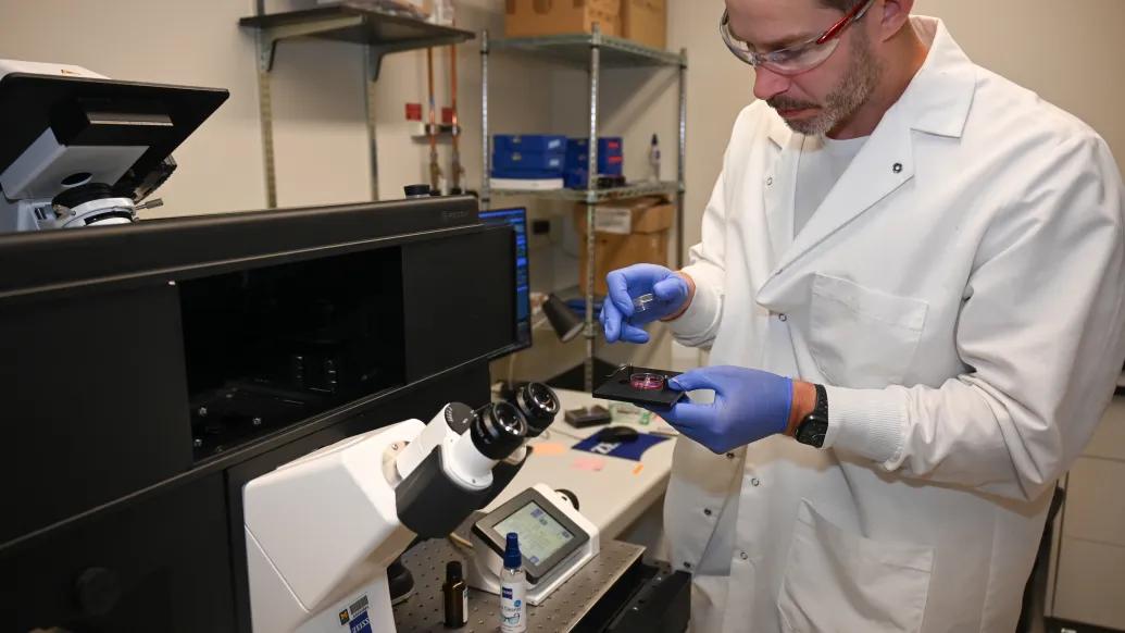
New microscope technology at Michigan Medicine is helping emergency medicine researchers unlock the brain’s most delicate secrets.
Thanks to a $1.2 million Department of Defense grant, scientists in the Department of Emergency Medicine can now study cells too fragile to image previously — pushing brain injury research forward with two advanced microscopes and a state-of-the-art brain monitor.
“We do a lot of translational research, and we needed a couple of instruments that do different things,” said Joe Wider, Ph.D., Research Assistant Professor of Emergency Medicine. “The DURIP program supports real, cutting-edge, but otherwise unaffordable technology.”
The grant supports tools that enable researchers to study injury at a molecular level, particularly traumatic brain injury (TBI), stroke, and cardiac arrest. Funded through the Defense University Research Instrumentation Program (DURIP), the grant was awarded to Dr. Wider; Thomas Sanderson, PhD; Hakam Tiba, MD, MS; and Cindy Hsu, MD, PhD, MS.
A Slide Scanner That Works While You Sleep
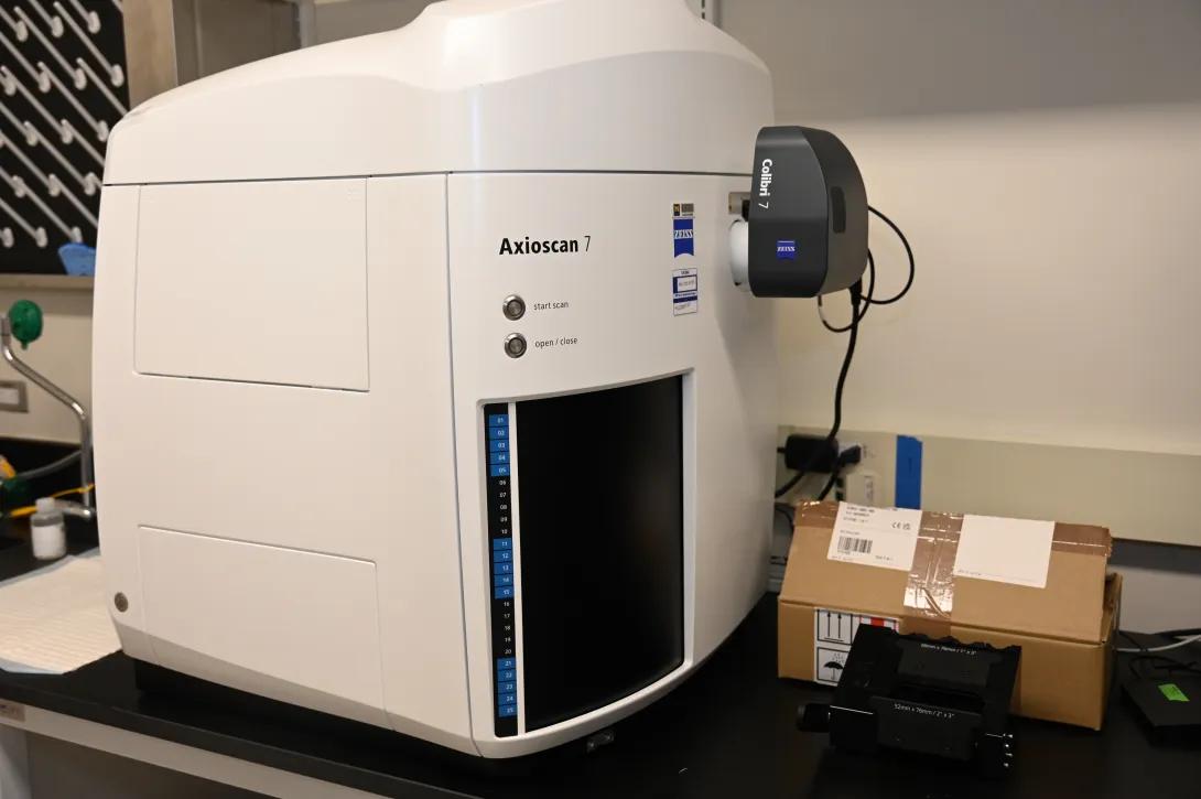
One of the biggest game changers is the Zeiss Axioscan 7, a fully automated slide-scanning microscope that can digitize 100 brain slides in a single run.
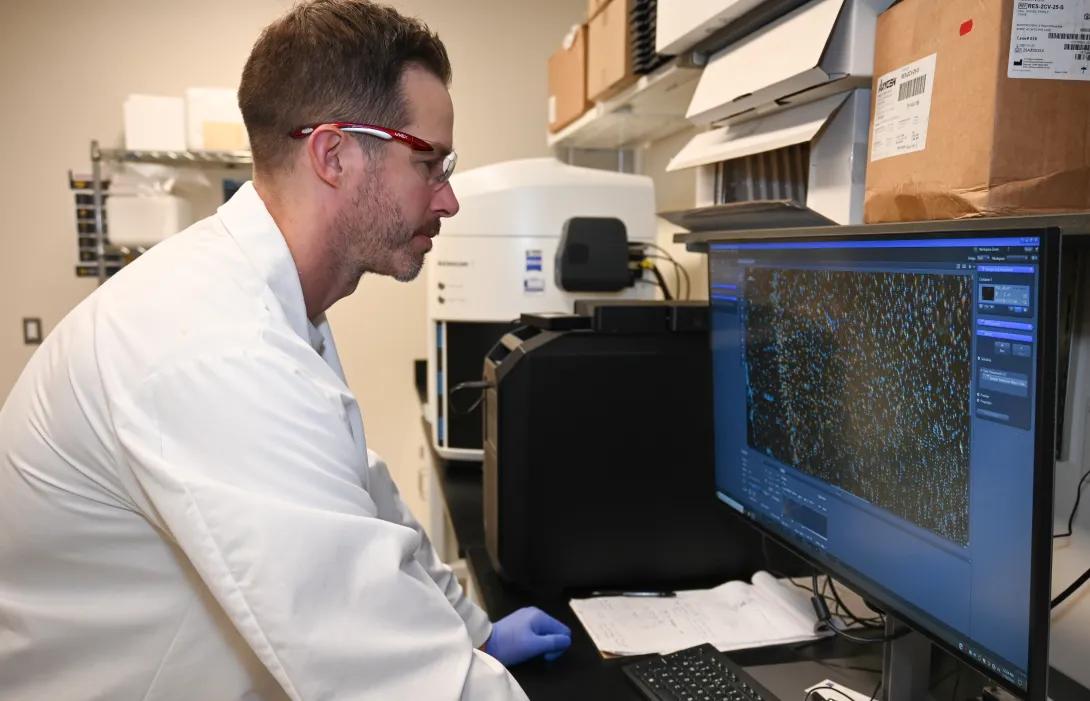
“We can image 50 pig slides back-to-back without changing anything,” Dr. Wider explained. “We just set it overnight, and it runs. Depending on the imaging we do, it might take an hour per slide, so it’ll run for 50 hours straight without anybody touching it.”
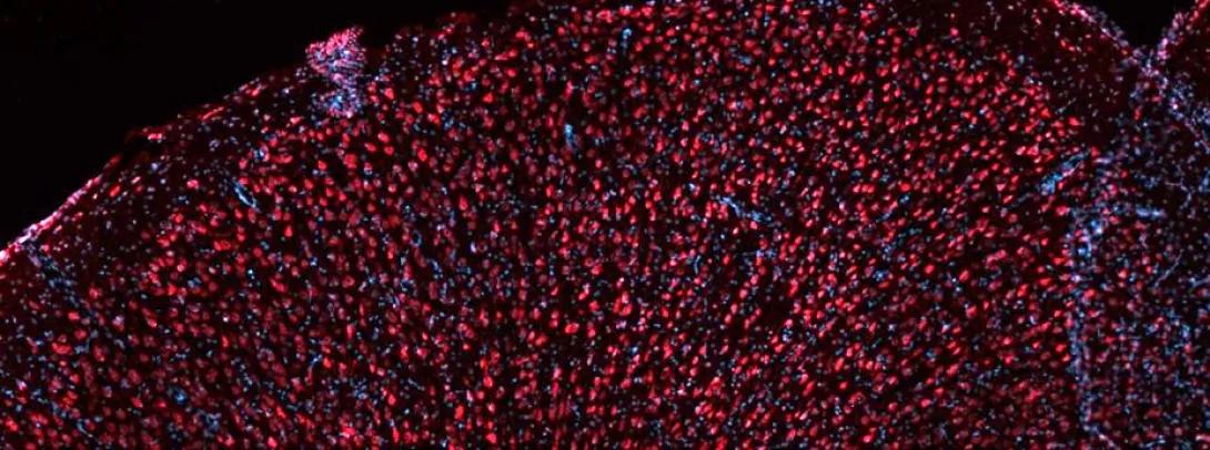
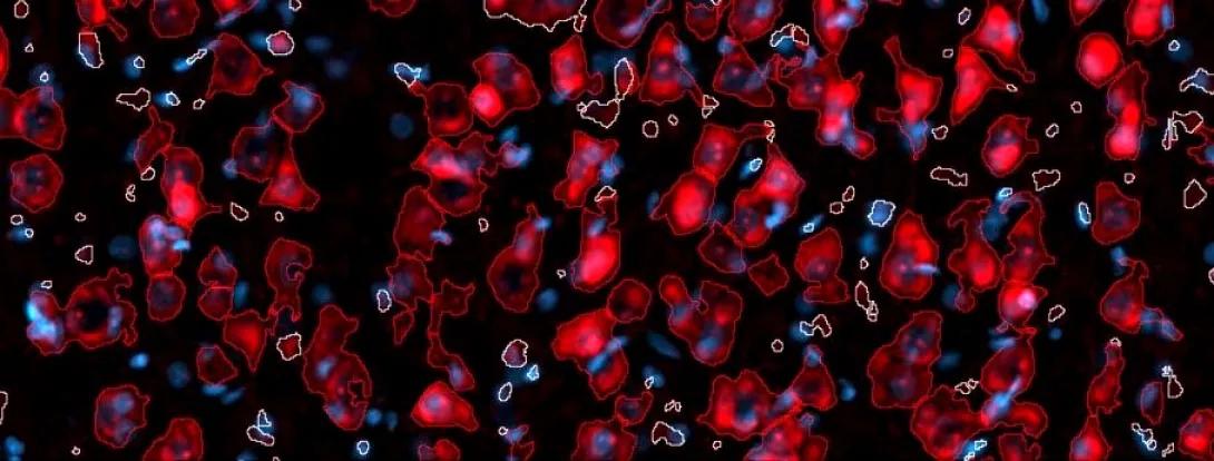
The scanner uses automated tissue detection, topographical mapping and AI-powered software to produce high-resolution images, which researchers analyze for changes in brain structure, like cell density and morphology.
These scans support a wide range of brain-injury research, including TBI, stroke, and cardiac arrest, led by both Dr. Wider’s lab and collaborators like Dr. Hsu.
“It’s been running basically 24/7 since we got it,” said Dr. Wider. “It’s been really useful.”
Seeing More, Using Less Light
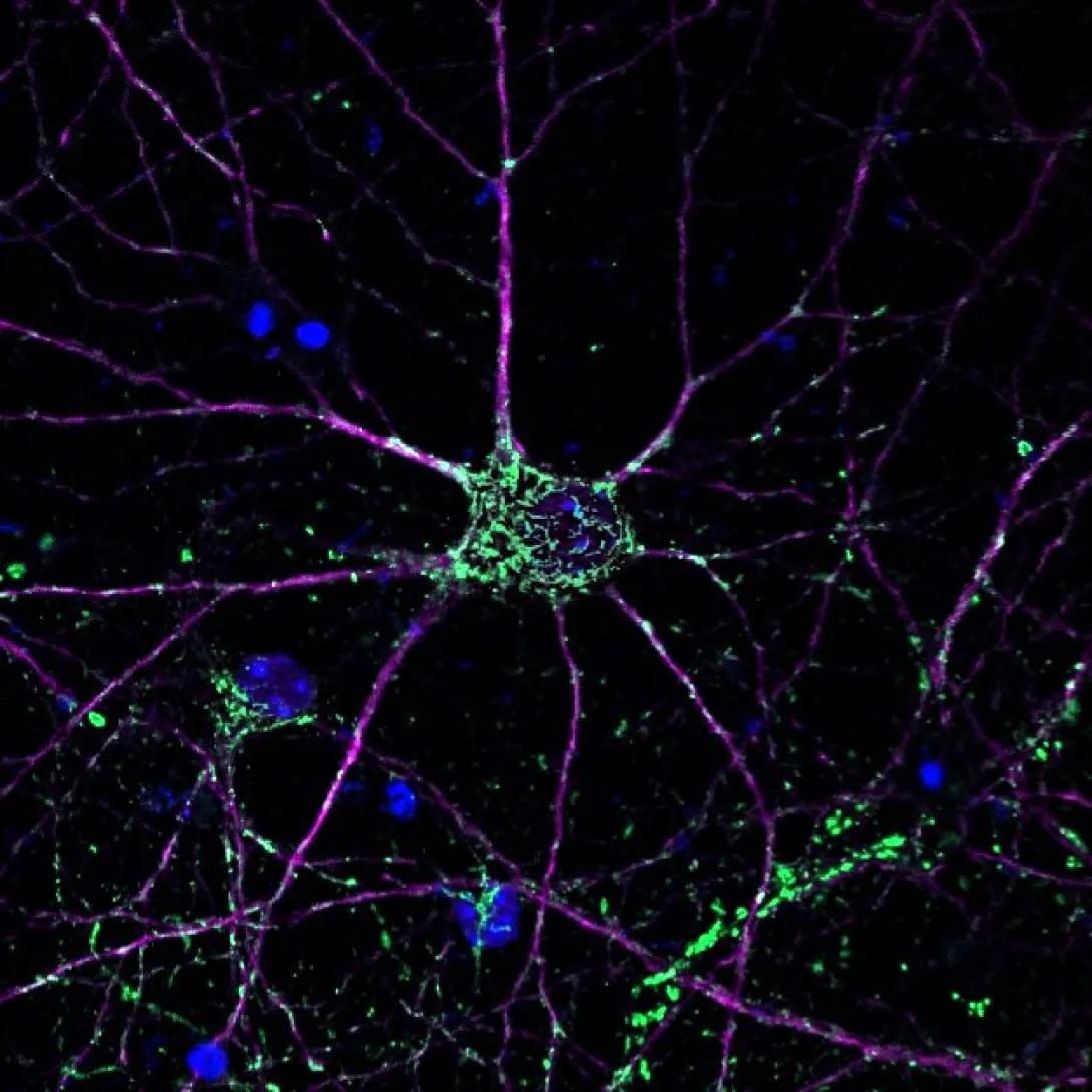
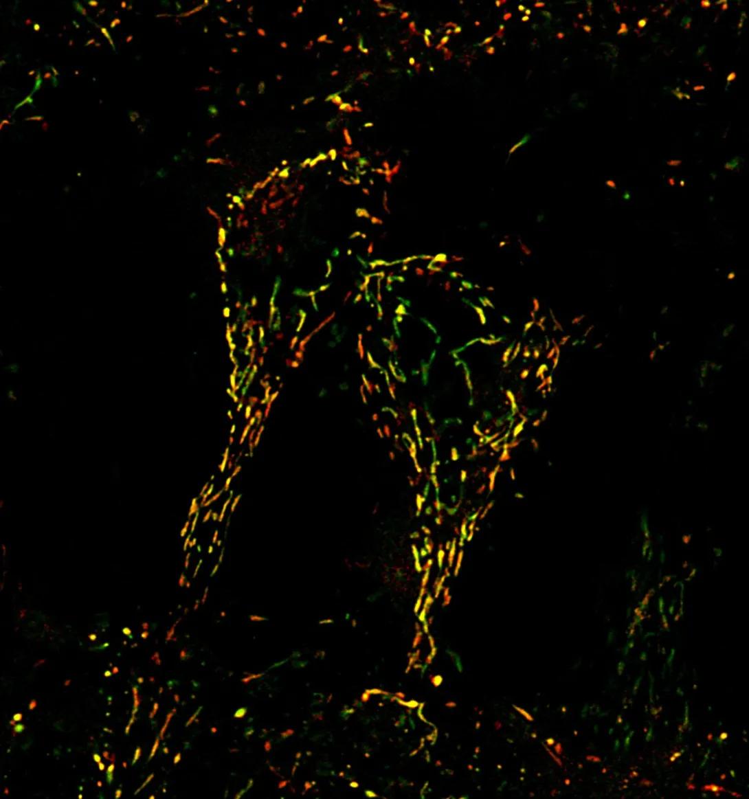
The lab also acquired the Zeiss LSM980 with Airyscan 2, a super-resolution microscope used for molecular and mechanistic research. Traditional confocal microscopy increases the signal-to-noise ratio and resolution by collecting light through a pinhole, which blocks out-of-focus light from reaching the detector. Airyscan technology utilizes an array of 32 light detector elements, rather than the traditional pinhole, to collect and computationally reconstruct the fluorescent signal into an image. In addition to improving resolution, this reduces the imaging time and light intensity.
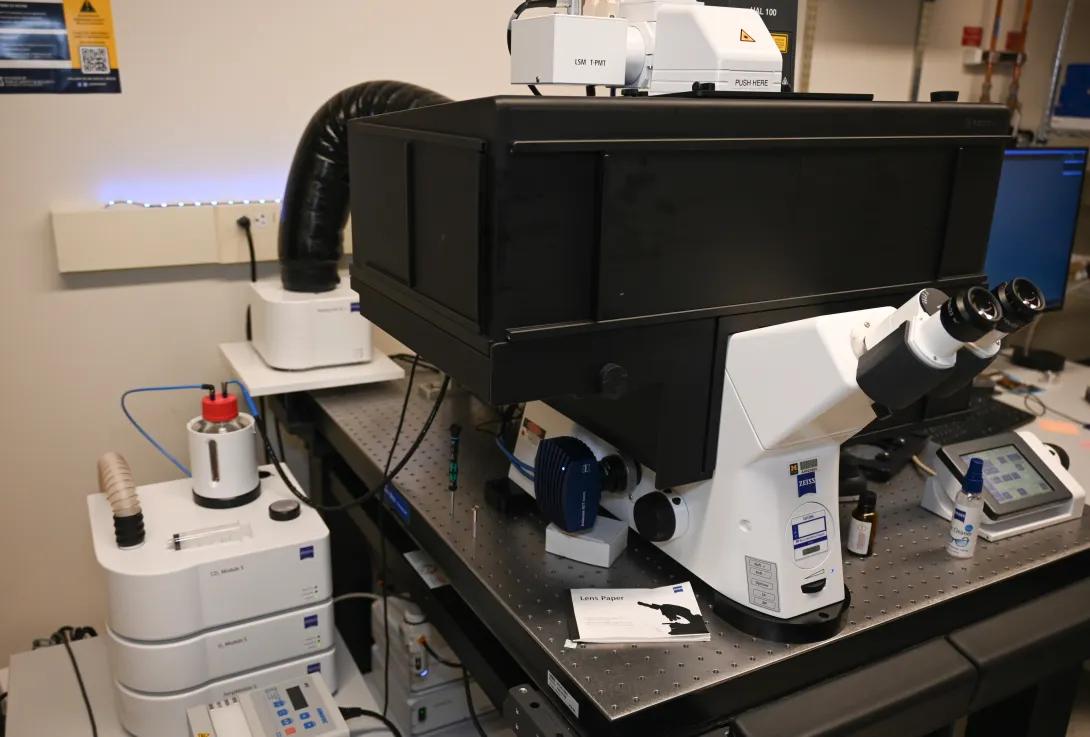
“That really reduces the amount of light that you’re shining on your sample,” Dr. Wider said. “It’s really nice for live-cell imaging, where a lot of light is toxic to your cells.”
Airyscan technology doubles the resolution compared to traditional confocal microscopy, to 100-120 nanometers. High magnification is essential for revealing subcellular detail.
“We can observe suborganellar structure,” Dr. Wider said.
Dr. Wider’s team is using the new instrument to track mitochondrial changes in models of TBI and cerebral ischemia. Super-resolution microscopy is necessary to see changes in mitochondrial ultrastructures — like the inner mitochondrial membrane cristae — which will offer new insight into how brain damage unfolds.
“We can get super-resolution imaging in live cells, without the cells dying,” Dr. Wider said. “This helps collect information over time about changes that occur in the cell under normal conditions and in response to stresses, like hypoxia and mechanical strain, with high temporal resolution.”
A Brain Monitor for Translational Models
The third tool, the Raumedic NEUROVENT-PTO, is an FDA-approved monitor that tracks brain tissue oxygenation, temperature, and intracranial pressure — all with one, minimally invasive probe.
“It’s cutting edge,” Dr. Wider said. “We’re using it in our translational models, and it’s producing a lot of good data. This is a neuromonitoring instrument used in the hospital setting, which contributes clinical relevance to our studies.”
The NEUROVENT is already in use with other teams, including Dr. Hakam Tiba’s group, which recently submitted a grant to expand translational research with it.
From Bench to Bedside
While still in the lab phase, the equipment has clear potential for clinical impact.
“We’re looking at the molecular mechanisms of brain injury, and these instruments help us discover new technologies and validate them in translational models,” Dr. Wider said. “It’s sort of from the bench to just before bedside.”
The new tools have already helped the team land new NIH funding.
“We were recently awarded a new R01 from NINDS to study molecular mechanisms of neurotrauma, and these instruments helped collect data
for our proposal,” Dr. Wider said.
Ultimately, Dr. Wider hopes this work leads to better diagnostics and treatment.
“Hopefully, we’ll be able to develop new technologies that can be used to improve patient outcomes,” he said.
Powering Progress, and the People Behind It
Beyond research, the new tools are also training the next generation. Students and postdocs from across the department are gaining hands-on experience.
“We’ve had a lot of students come through learning to use this equipment,” Dr. Wider said.
This progress also builds on years of support from the Weil Institute for Critical Care Research and Innovation.
“A lot of the preliminary data used in the proposal was supported by the Massey Grand Challenge, which is a Weil Institute initiative,” Dr. Wider said. “They funded the early research that led to this instrumentation program.”
From scanning hundreds of slides overnight to imaging live neurons at nanoscale resolution, these tools are already accelerating discovery.
“It’s been a lot of fun using the equipment,” Dr. Wider said. “The labs in Emergency Medicine have lot of new and ongoing studies, including research funded by the Massey Grand Challenge, the Kahn Pediatric Critical Care Grand Challenge, and NINDS. And we are always collecting data for new proposals. So, this is just the beginning — we expect these instruments to get a lot of use.”
In This Story
Joseph Wider, PhD
Assistant Professor
Thomas Sanderson, PhD
Professor
Mohamad Hakam Tiba
Research Associate Professor
Cindy H Hsu, MD, PhD, MS
Associate Professor
Featured News & Stories
Nerve blocks for kids’ broken legs: U-M helps show a safer, faster way to stop the pain
Medicaid billed for 52% of U.S. hospital costs from gun injuries
Could ketamine be the key to stopping life-threatening seizures?
The future, fast: Emergency Medicine at U-M positions AI as a department-wide strategy
A new way to fight sepsis