3D-Printed Models: Conjoined Twins
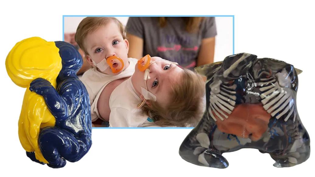
The 3D-printed models featured in a year-long Detroit Metropolitan Airport (DTW) exhibition show how Michigan Medicine teams used advanced medical imaging, 3D modeling, and hands-on surgical planning to prepare for the successful separation of conjoined twins.
Conjoined twins are identical twins who do not fully separate early in pregnancy. They may be born physically connected and, in some cases, share organs or other internal structures. Conjoined twinning is very rare, estimated to occur in roughly 1 in 50,000 to 1 in 200,000 births. Surgical separation is possible in some cases, depending on the anatomy involved and which structures are shared.
For this complex case, Michigan Medicine physicians and imaging specialists created life-size models before and after delivery. The models helped care teams better understand the twins’ anatomy, plan the safest surgical approach and rehearse important steps before the procedure.
Why 3D Models Matter
When anatomy is especially complex, a physical model can help clinical teams see details that are difficult to fully understand on a screen. These models gave surgeons, radiologists, anesthesiologists, nurses and other specialists a shared reference point as they planned the twins’ care.
The models helped the team:
- Understand which structures were shared and which were separate
- Plan the safest approach for separation
- Practice handling, transfer and surgical steps
- Communicate across multiple specialties involved in the twins’ care
- Educate the family
Having a life-size model let our team rehearse key steps and anticipate challenges before we ever entered the operating room. The separation was a success.”
Dr. George Mychaliska, C. S. Mott Children's Hospital
Explore the 3D Models
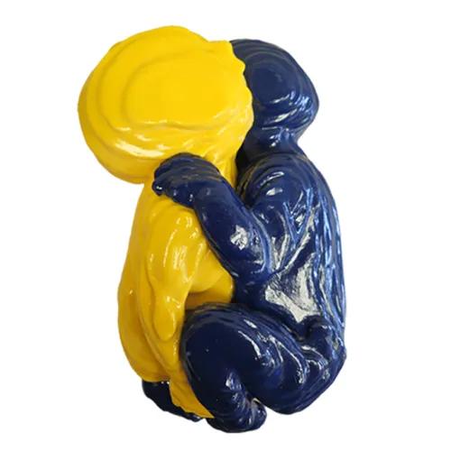
Maize & Blue Model
Created from an MRI scan before delivery, this model helped the team prepare before the twins were born.
The twins appear to be “hugging”, but they are connected to each other.
Other MRI images show the connection between them.
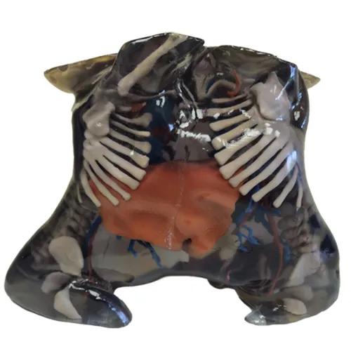
Transparent Model
Created from a CT scan after delivery, this model shows the connection between the two girls from the chest to the pelvis.
The skin and muscles were printed with transparent material to show internal structures, including the shared liver (orange), bones (tan), arteries (red) and veins (blue).
Credits
The models were created by William Weadock, M.D., and members of the U-M Medical School Department of Radiology 3D Printing Imaging Laboratory.
The models were printed at the U-M Duderstadt Center Fabrication Lab.
In This Story

William J Weadock, MD, FACR
Clinical Professor
Related

Michigan Medicine Team Separates Conjoined Twins at C.S. Mott Children’s Hospital
Featured News & Stories

Learning about the Life Loaf: My Experience Working in OBGYN Research

What’s the latest on 7-OH and kratom availability and addiction care?

MDisability and CDHW interns present their final research presentations

New oral nanomedicine boosts effectiveness of cancer immunotherapies

A first-of-its-kind treatment for mastocytosis