Cultivating a Passion for Ultrasound: How it Began with Diagnostics and Therapeutics
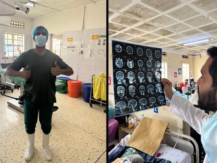
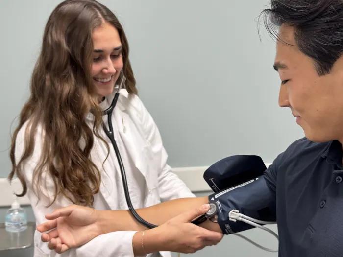
I placed a generous amount of ultrasound gel onto a curvilinear transducer and watched white, gray and black flash onto the screen as I pressed on the abdomen of the human ultrasound model. What started out as indistinguishable bright and dark structures soon became the abdominal aorta, the inferior vena cava, loops of bowel and vertebrae. Just earlier that week, we had learned how to find free fluid using a Focused Assessment with Sonography for Trauma (FAST) exam, a diagnostic procedure that could save a patient’s life after a trauma by localizing organ bleeding. Later, we learned how to visualize stones inside or free fluid around the gallbladder, perform an echocardiogram to assess the heart, diagnose air or fluid around the lung and perform various procedural interventions, all with the use of ultrasound imaging. One afternoon, a team of my peers and I employed all that we learned to perform diagnostic scans on real patients in the Department of Emergency Medicine with the guidance of a faculty member or fellow.
This is all a part of the beginning of M3 year, known as Branch Launch, when we participate in one of four special “Branches” after we are done with the Scientific and Clinical “Trunk.” I participated in the newly restructured Diagnostics and Therapeutics (D&T) Branch which turned out to be an incredible opportunity to have intensive exposure to ultrasound. Before each day of hands-on learning with real people as ultrasound models, we watched easily digestible, 5-minute videos from Core Ultrasound. Faculty and residents demonstrated how to find structures and guided us during the ample time we had with transducers in our hands. Groups of students did not exceed three per model, which afforded enough time for us to scan until we felt confident in our abilities to evaluate anatomical structures.
Lectures included a wide variety of topics such as ultrasound use in the eye, 3D-printing based on CT imaging and endoscopic surgery management of obesity. All lectures had a component of imaging and/or therapeutics which is distinct from most of our medical education leading up to this course. D&T offers a unique setting to be exposed to faculty, fellows and residents interested in the clinical use of ultrasound earlier in our education. One of the most beneficial aspects of this Branch was gaining wonderful mentors organically through teaching done in small groups.
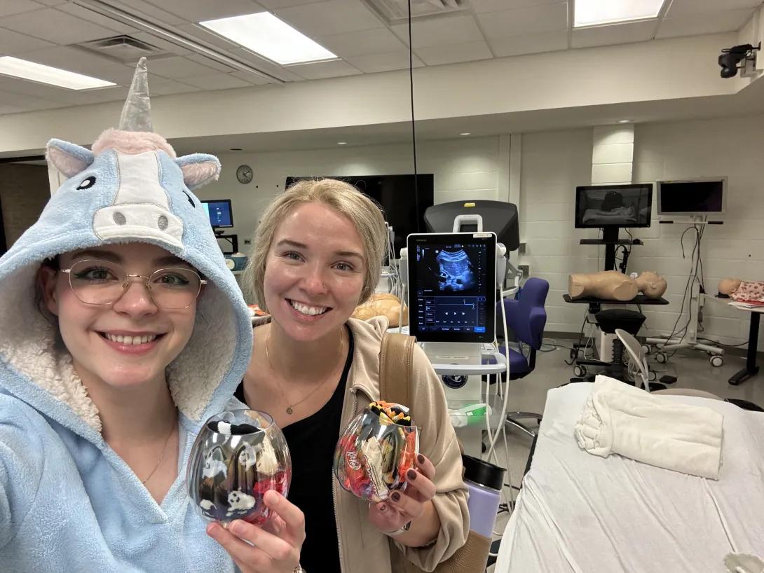
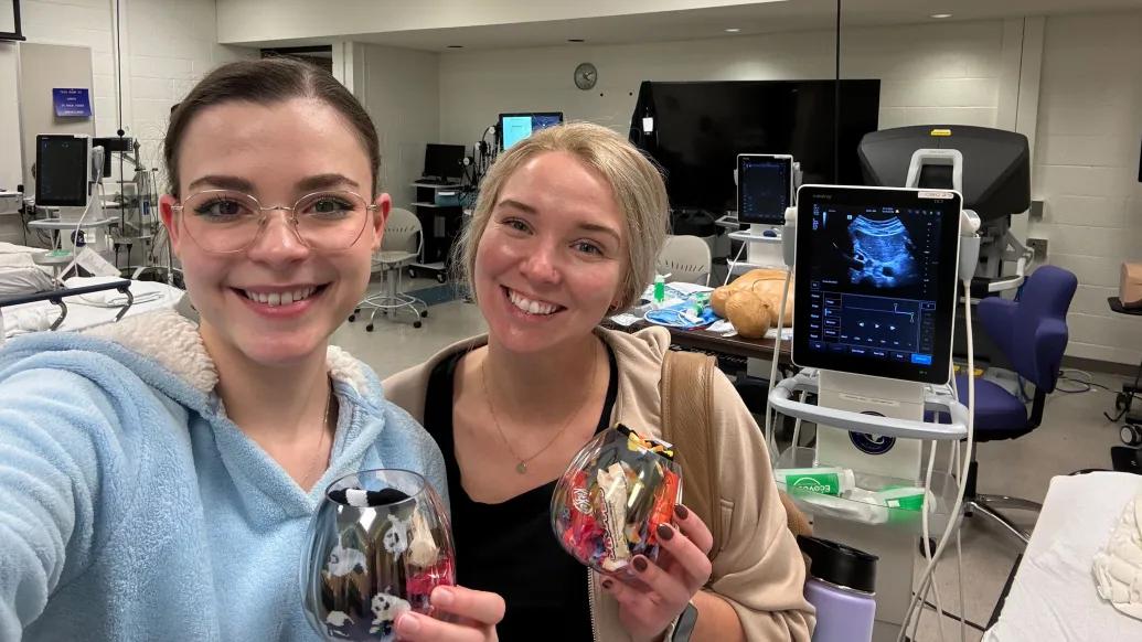
At the end of two weeks dedicated to D&T, we put on our Halloween costumes and participated in Simulation Games Day. Five teams of 5-6 students each competed to be the best at ultrasound scanning and trivia. My team ended up winning second place! My unicorn costume must have contributed to our luck in identifying structures and answering questions swiftly. The competition day was a fun way to demonstrate an explosion of skill learned in just two weeks.
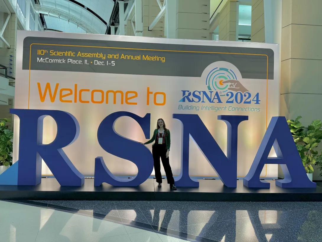
One month later, I took my interest in ultrasound to the next level by attending specialty hands-on labs offered at the Radiological Society of North America (RSNA) conference in Chicago, Illinois. The three courses I attended elaborated on scanning methods for pediatric musculoskeletal, foot and ankle, and carotid and abdominal vasculature. I deepened my skills with the help of supervising radiologists from all over the United States, Argentina, Chile and Brazil. It was a privilege to scan live models amongst faculty and residents from all over the world (including Germany, France and the mentioned South American countries). What had begun as curiosity with ultrasound in my M1 year transformed into fascination during the D&T Branch. With further exploration, it has become a passion that I hope to expand upon with research in my future career.
For those who did not participate in D&T or who plan to participate in a different Branch and still want ultrasound exposure, I recommend taking the 2-week ultrasound elective through the Department of Anesthesiology. Matt Sparling, a M4 and future anesthesiologist, constructed this excellent self-paced and asynchronous course that utilizes a mannequin simulator to teach basic ultrasound skills. Moving forward, Matt will be transitioning the management of the course to me, and I am excited to help you in the pursuit of learning ultrasound!
Rigorous use of ultrasound is certainly not limited to radiology. In fact, this technology unites specialties across our hospital. Anesthesiology, critical care and emergency medicine heavily utilize ultrasound for procedures including vascular access and nerve blocks. Point-of-care ultrasound (POCUS) is becoming the standard in emergency medicine and pediatrics due to rapid data collection and avoidance of ionizing radiation. Obstetrics and gynecology and surgery leverage diagnostic data to make clinical decisions daily. Internal medicine and family medicine physicians are becoming exceedingly interested in learning how to leverage ultrasound to better care for their patients. More focused paths are available and include ultrasound fellowship after emergency medicine or family medicine residency. This is certainly a tool that has become and will continue to be ubiquitous in many specialties.
What started as the best Branch option transformed into a career interest in ultrasound for me. My experience in D&T is one of my favorite memories in medical school because of the passion it cultivated in ultrasound and the excellent teaching we received. Ultrasound is a modality many of us future physicians will need to learn as it becomes increasingly essential to patient care. Thank you to Dr. Kate Klein, Dr. Will Kropf, Dr. Eve Losman, Dr. Alisha Ching and Angela Hickey for making D&T such a wonderful course!

Melis Ozkan is a third-year medical student at the University of Michigan Medical School. In her free time, she likes to dance and choreograph for Biorhythms and The Smoker. She can be added on LinkedIn here and on Instagram here.
Media Contact
MD Admissions
University of Michigan Medical School
In This Story
Katherine A Klein, MD, FACR
Clinical Professor
Charles "Will" Kropf
Clinical Assistant Professor
Eve Losman, MD
Clinical Associate Professor
Featured News & Stories

Thank You For This Interesting Consult: Preparing for Residency with Friendly Competition
Love Is Our Resistance: Redefining Advocacy in the Patients & Populations Branch
Our Journey with the Doctoring Course
From Studying to Scrubs: Preparing for M2 Year with Transition to Clerkships
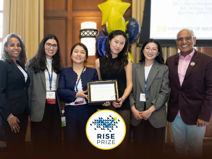
Inaugural RISE Prize Recognizes Innovation in Health Sciences Education